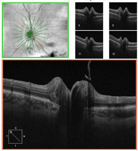
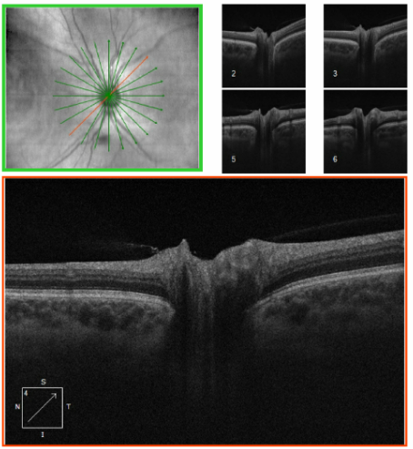
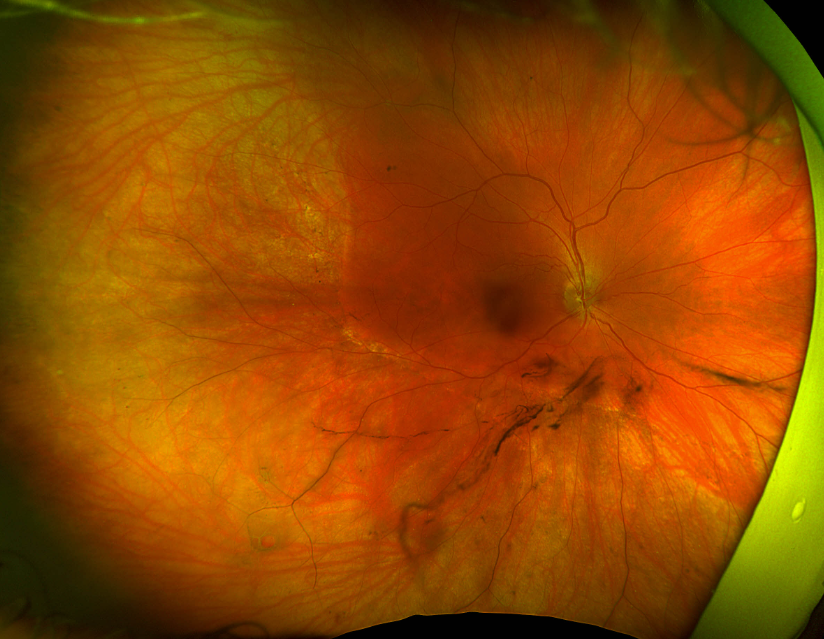
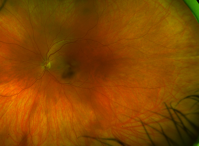
Yes, I agree that the discs don't look all that impressive in the optos photos, but there is a clear asymmetry clinically, with the right disc being more elevated and hyperemic compared to the left. I thought the 10um difference and superior thickening on OCT confirmed my clinical impression.
As for EDI OCT, our Heidelberg is currently down, so I got the deepest images I could on the Cirrus, but I feel confident about the lack of ODD (our study with Steffen showed that FAF is still pretty darn good). I did try to capture the one slice that showed the little papillary tuft, although I'm not convinced that it's enough (or in the right location) to be the cause.
For the surgical approach, a buckle without vitrectomy is the plan, since she is so young and so myopic. If she gets a post-vitrectomy cataract in that eye, she will not be able to tolerate the anisometropia if we fix it, which opens up a whole new set of issues.
I think/hope I've answered all the questions.. As always, I appreciate everyone's input!
Lulu
The information transmitted is intended only for the person or entity to which it is addressed and may contain confidential and/or privileged material. Any review, retransmission, dissemination or other use of, or taking of any action in reliance upon, this information by persons or entities other than the intended recipient is prohibited. If you received this in error, please contact the sender and delete the material from any computer.
www.sjhc.london.on.ca
Original Message:
Sent: 7/10/2025 7:16:00 PM
From: Eric Singman
Subject: RE: Retinal detachment and optic disc edema
Where do we stand on PVD and NAION? Possible "mild" case. VEP?
Original Message:
Sent: 7/10/2025 6:38:00 PM
From: Christine Greer
Subject: RE: Retinal detachment and optic disc edema
Hi!
I bet a PVD caused both a tear & detachment and also caused some traction at the disc as the vit separated off the optic nerve head. Is these a Weiss ring? I’ve seen real time disc edema and hemorrhage occur during RD repair when the surgeon is inducing a PVD. It’s wild.
Wouldn’t stop the surgeon from doing a buckle if that’s what’s indicated from a retinal perspective.
Christine
Original Message:
Sent: 7/10/2025 5:22:00 PM
From: Matthew Kay
Subject: RE: Retinal detachment and optic disc edema
If these are the photos of the patient that was described as having optic nerve edema with a retinal detachment, these optic nerves do not appear to be swollen in the least.
I would do absolutely nothing from a neuro-ophth perspective.
Matt
Sent via the Samsung Galaxy S21 5G, an AT&T 5G smartphone
Original Message:
Sent: 7/10/2025 1:41:00 PM
From: Lulu Bursztyn
Subject: RE: Retinal detachment and optic disc edema
Thanks everyone for your input, and an interesting debate on Occam's razor...
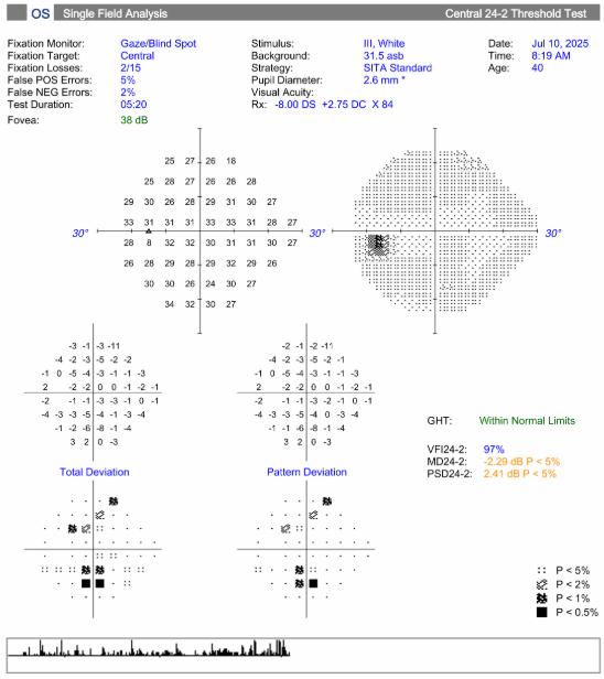
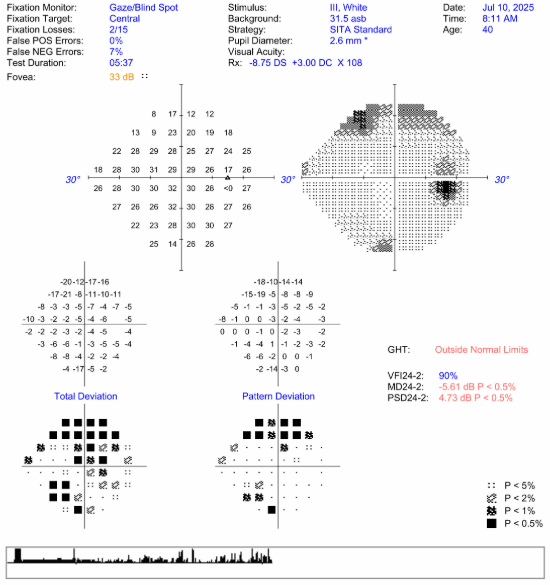
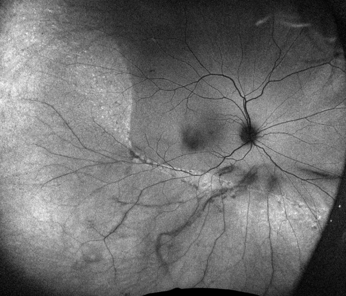
Some updates, now that I've had a better look at her, and hopefully the images come through. She describes new floaters starting one week ago followed by a superior curtain defect over the past few days. Prior to this vision was her normal. Visual acuity remains 20/80 R, 20/20 left, colour 16/16 OU. IOP was 15 OU this morning. I actually cannot detect an RAPD despite the complaint of subjective decreased brightness. Anterior chamber is quiet, no vitritis, just a lot of vitreous syneresis (this is what I mean by turbid – very thick strands of floaters). There is an inferior temporal hole in the RD. There is bilateral fairly symmetric inferior RNFL thinning that I suspect is related to her high myopia, but there is also superonasal edema of the right disc. The visual fields show a superior field defect in the right as well as a small inferior defect in both eyes. I have requested an MRI (this is Canada, it's not happening today...) and have sent her for some blood work (CBC, CRP, syphilis, ENA, mitochondrial panel...). I will also see her back after the RD is fixed.
Lulu

Hole visible inferotemporal
FAF, no visible ODD, but the RD really stands out nicely
Lulu Bursztyn
Comprehensive and neuro-ophthalmology
Western University, London, Ontario
lulu.bursztyn@sjhc.london.on.ca
519-646-6214
The information transmitted is intended only for the person or entity to which it is addressed and may contain confidential and/or privileged material. Any review, retransmission, dissemination or other use of, or taking of any action in reliance upon, this information by persons or entities other than the intended recipient is prohibited. If you received this in error, please contact the sender and delete the material from any computer.
www.sjhc.london.on.ca
Original Message:
Sent: 7/10/2025 11:27:00 AM
From: Sherif Ahmed Kamel Abdelbar
Subject: RE: Retinal detachment and optic disc edema
Things are not so simple especially when it comes to neuro-ophthalmology or retinal problems , then combined proposed simplicity may be a walking door to problems
Original Message:
Sent: 7/10/2025 11:14:00 AM
From: Scott Forman
Subject: RE: Retinal detachment and optic disc edema
I think Occam's razor is an outdated concept developed before medicine got as complex as it is. Multiple problems (not necessarily totally unrelated) are common and yes they occur in the same body but many are handled by different approaches.
+=+=+=+=+=+=+=+=+=+=+=+=+=+=+=+=+=+=+=+=
Scott Forman, MD
Senior Fellow North American Neuro-ophthalmology Society
Adult and Pediatric Neuro-ophthalmology
Comprehensive Ophthalmology
Functional Medicine