NANOSNET

 View Only
  • 1.  Scotoma, Patchy Choroidal Filling and Retinal vein outflow abnormalities

    Posted 2 days ago
    Edited by Erin Lanzo 2 days ago

    Hi Everyone,

    Long time listener, first time caller here. Thanks so much in advance.

    My case is a 50 yo male with PMH OSA, Meniere's, obesity (on GLP1), HLD who on July 4 noted a nasal scotoma in his right eye after doing water sports in Europe (diving and "under water jet skiing' on a "Seabob"). 

    On 7/28 - saw his general ophthalmologist who noted this, but an otherwise "normal" exam with 20/20 central vision.
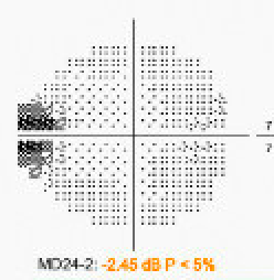
    He had retina follow up for further testing on 8/3. An FA was done showing patchy choroidal filling OD and delayed filling of a superior retinal vein. Below are two stills - unfortunately there are no time stamps. Incidentally noted at this retina appointment was an inferotemporal shallow RD in the left eye, not causing any visual symptoms. Still 20/20 centrally OU. IOP wnl, no cells or signs of inflammation otherwise in AC or vit.
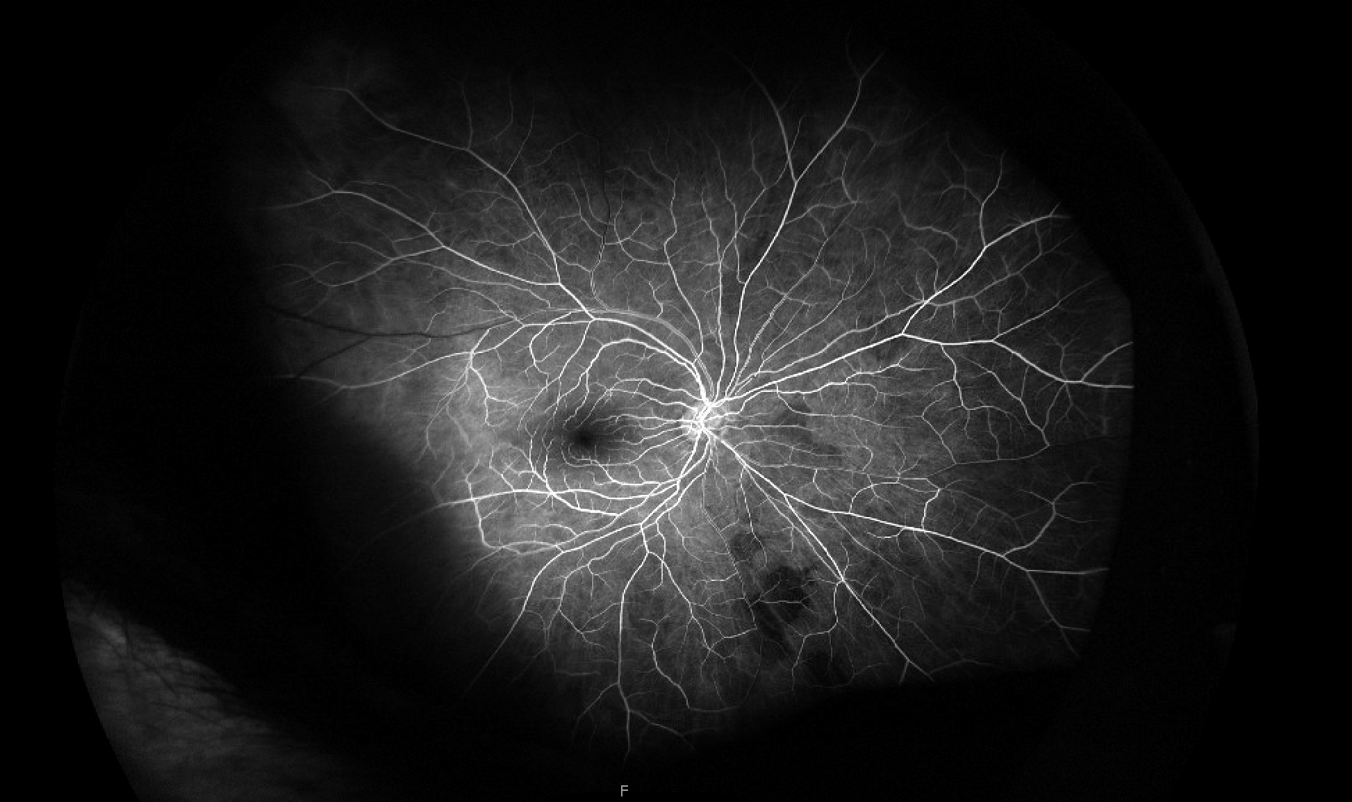
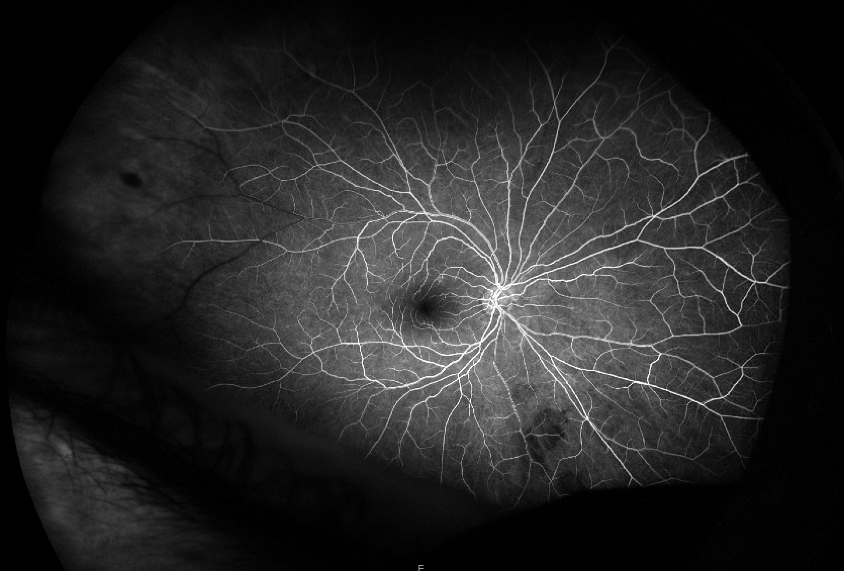
    The retina MD was understandably concerned about the patchy choroidal filling and sent him to ED for OIS work up.

    MRI Brain and orbit w/wo was normal (I also reviewed - no perineural or optic nerve enhancement, no orbital fat enhancement), CTA H/N showed no stenosis and ESR and CRP were both <1. GCA ROS negative except for headaches off and on over the month, unchanged in quality compared to his "usual" headaches (bifrontal, retroorbital). He has had some dizziness spells in July which are similar to prior spells he associates with his Meniere's disease, but perhaps have occurred more frequently in past couple of weeks. He had a lone star tick bite in June but does not have systemic symptoms/signs of tick borne illness otherwise (no fevers, no rash). He also reports having had a few "flashes" OD.

    When I saw him 8/4 - scotoma was similar if not a bit less dense on my HVF. He denied any episodes of amaurosis leading up to our visit.

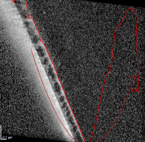
    I noted that there was some peripheral schisis temporally OD that may correspond with his scotoma, though per discussion with retina, this looks like normal anatomic peripheral schisis. The scotoma seems to correspond with the area in the temporal macula/midperiphery where the retinal vein has delayed filling, but there are no other signs of BRVO. No other retinal or nerve signs of ischemia either (no PAMM, no disc edema, no RAO etc).

    Here's the best quality OCT I could get through that area of temporal retina. Mac OCT was normal.
    I sent a broader hypercoaguablity and inflammatory work up in addition to some labs that neurology sent from the hospital. Thus far:

    Negative/normal: ESR <1, CRP <1, CBC,  plts 211, RF, ANCA screening, Lyme, SPEP, homocysteine, fibrinogen, lupus anticoagulant, protein S&C, ATIII activity, APTT, PT/INR, folate, B12, trep Ab, ANA screen,

    Abnormal: A1c 5.9, high thrombin time (63, normal <25).

    Pending:  Rickettsia (other tick screen PCR was negative), quant, lysozyme, ACE, an echo

    He's seeing retina again next week -- the shallow RD in the left eye was lasered and did appear to them to be a true RD rather than schisis, but they plan to re-evaluate this prior to surgery given what's going on with the right eye. 

    My main questions are:

    -is there something I'm missing with respect to the patchy choroidal filling in terms of work up? I have low suspicion for GCA base on young age, negative inflammatory markers and ROS, as well as (thus far) lack of amaurosis. and other signs of ischemia. Would you nonetheless do a biopsy in this patient? He is not eager to do so. 

    -is there a unifying pathology that would cause patchy choroidal filling and focal delayed venous outflow? 

    Thanks so much for your thoughts.

    Erin


  • 2.  RE: Scotoma, Patchy Choroidal Filling and Retinal vein outflow abnormalities

    Posted 2 days ago
    1) Patchy choroidal filling is common in patients over age 50, seen in at least 25% of patients with AMD in the CATT trial. This is a red herring. It is not a sign of GCA or ocular ischemic disease. Large choroidal non-perfusion is what has been reported in GCA.

    2) how far posterior does the schisis stretch? there appears to be reduced vascular density in the superotemporal perimacula.

    Screenshot 2026-08-06 at 3.00.29 PM.png
    Is there sup-temporal macular thinning on the volume map of OCT macula compared to infero-temporal?

    2) Are there early phase (Arterial) of the fluorescein? I agree it doesn't look like BRVO, there is limited arteriolar perfusion supero-temporally, this may be reperfused / old BRAO or small vessel ischemia (sickle trait is probably the most common cause of small vessel ischemia without acute onset). Do you have macular OCTA to see if there is macular non-perfusion?

    Best,

    Drew


  • 3.  RE: Scotoma, Patchy Choroidal Filling and Retinal vein outflow abnormalities

    Posted 2 days ago
    Edited by Erin Lanzo 2 days ago

    Thank you! 
    Here are earlier sequential arterial phase stills (again, no timestamps available)

    ^of note, this vein does eventually fill. 

    The mac cube volumetric data shows maybe some asymmetry between OS and OD, superiorly mainly, but the area you've highlighted may be just outside the border of the cube:

    and lastly -- really tough to say the extent of the schisis OD. I honestly could not appreciate it on my fundus exam but was prompted to do the OCT out in the temporal periphery because of the RD/schisis connundrum in OS. You can see a demarcation line in OS but not OD:

    No Mac OCTA yet - but easy for me to get. Thanks for this suggestion!


  • 4.  RE: Scotoma, Patchy Choroidal Filling and Retinal vein outflow abnormalities

    Posted 2 days ago
    Hi Erin, good case. 
    You may know this already, but more shallow defects are present with RD and full blackout/punched out defects are more classic with retinoschisis. That far inferotemporal in the usual schisis location that  I appreciate on the photos shouldn't cause a defect in a 24 degree field. 
    The field defect might be due to a small peripheral branchlet artery occlusion that reperfused, maybe triggered by the valsalva of diving/jet skiing.

    In terms of the choroidal filling patchy defects, I agree they are patchy up past AV phase, so that to me is concerning for ophthalmic artery poor perfusion. I agree with Drew that large choroidal defects are more characteristic of GCA, but the area your FA demonstrates in the early AV laminar flow phase is through the nerve/watershed zone that we do see in GCA with AAION-without disc edema, not as convinced it relates. In this age group I would not be worried about GCA anyway unless he had tons of systemic ROS+, but I wouldn't ignore the perfusion issue either so in my world full of vasculopaths, I would have gotten CTA head/neck and looked for carotid disease of significance to assess stroke risk. 

    - I would not do a TA Biopsy in this patient, not without something more than just FA changes. 
    - I would repeat the visual field/scotoma asessement in a month or two. If you can get a kinetic full field, it might be better to see what is going on with the schisis/RD. Otherwise, would see what the RNFL And macular GCL looks like in 3 mos and modify general vascular risk factors.

    @drew-super interested in the patchy choroidal ddx as we use this assessment to break the tie between doing/not doing a TAB even in patients without AAION. Why would this happen in AMD? And was is in areas separate from those of the geographic atrophy or choroidal disruption/NV?
    Kim


  • 5.  RE: Scotoma, Patchy Choroidal Filling and Retinal vein outflow abnormalities

    Posted 18 hours ago
    Good morning 
    Very interesting case from the story and the pictures we must do Carotid and transcranial Dopplex study may give some valuable information as well as cardiological consultation with recommending doing Echo heart 
    My recommendation by suspicion and you can say reasonable doubt is to shift to another drug from GLP , treat sleep apnea seriously , and stop diving and snorkelling for now at least 
    Two weeks ago I had a young women with what you can say suspicious retinal and optic nerve heat ischemia not obese no systemic risk factors all imaging is normal , she had sever sleep apnea and night stomach regurgitation 
    Another case in a young obese women with hyperlipidemia with the same complaints and what appears to be retinal and optic nerve ischemia , she was using GLP drug for months 
    In you case the retinal problem may cause some confusion but in the same time it may be origination from the same etiology , sleep apnea and diving , at least indirectly 
    Very intersecting case 
    Sherif