Hi Everyone,
Long time listener, first time caller here. Thanks so much in advance.
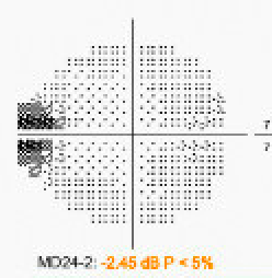
My case is a 50 yo male with PMH OSA, Meniere's, obesity (on GLP1), HLD who on July 4 noted a nasal scotoma in his right eye after doing water sports in Europe (diving and "under water jet skiing' on a "Seabob").
On 7/28 - saw his general ophthalmologist who noted this, but an otherwise "normal" exam with 20/20 central vision.
He had retina follow up for further testing on 8/3. An FA was done showing patchy choroidal filling OD and delayed filling of a superior retinal vein. Below are two stills - unfortunately there are no time stamps. Incidentally noted at this retina appointment was an inferotemporal shallow RD in the left eye, not causing any visual symptoms. Still 20/20 centrally OU. IOP wnl, no cells or signs of inflammation otherwise in AC or vit.
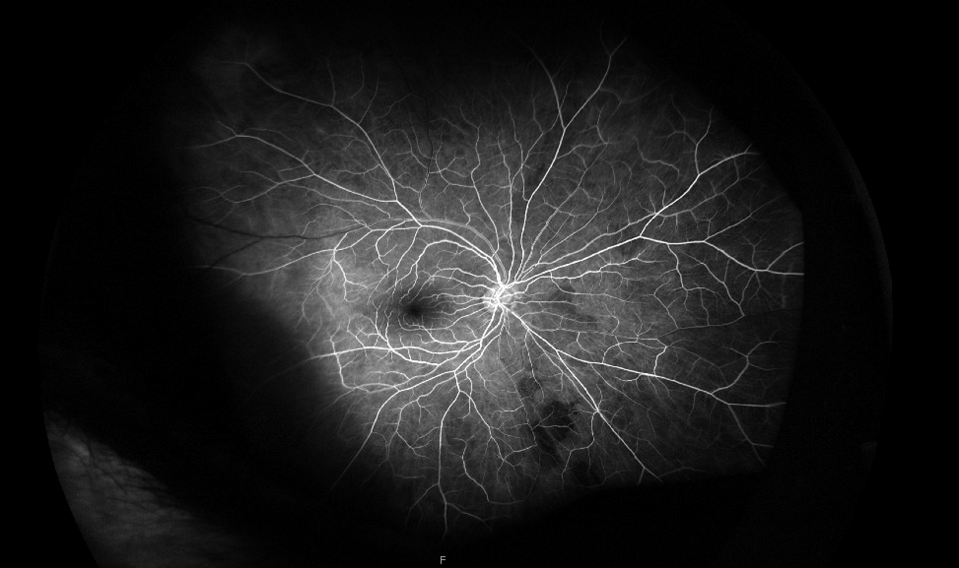
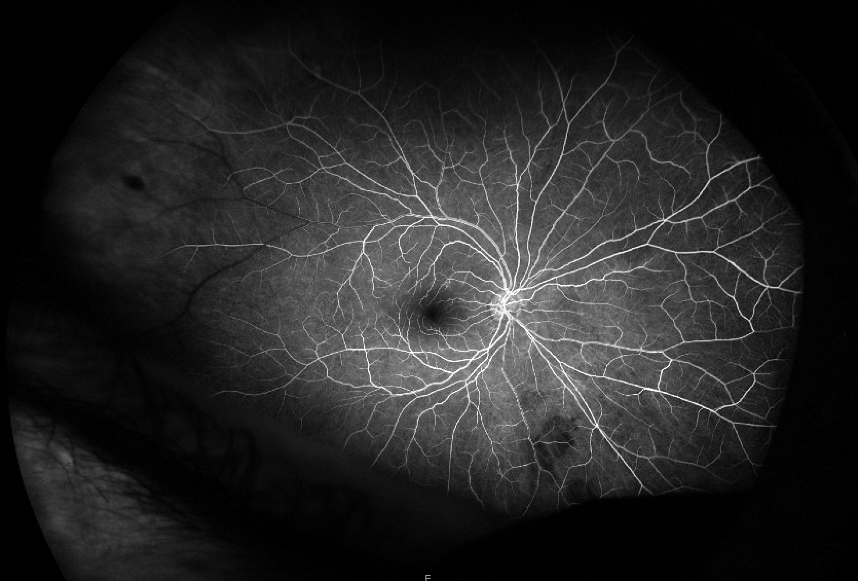
The retina MD was understandably concerned about the patchy choroidal filling and sent him to ED for OIS work up.
MRI Brain and orbit w/wo was normal (I also reviewed - no perineural or optic nerve enhancement, no orbital fat enhancement), CTA H/N showed no stenosis and ESR and CRP were both <1. GCA ROS negative except for headaches off and on over the month, unchanged in quality compared to his "usual" headaches (bifrontal, retroorbital). He has had some dizziness spells in July which are similar to prior spells he associates with his Meniere's disease, but perhaps have occurred more frequently in past couple of weeks. He had a lone star tick bite in June but does not have systemic symptoms/signs of tick borne illness otherwise (no fevers, no rash). He also reports having had a few "flashes" OD.
When I saw him 8/4 - scotoma was similar if not a bit less dense on my HVF. He denied any episodes of amaurosis leading up to our visit.
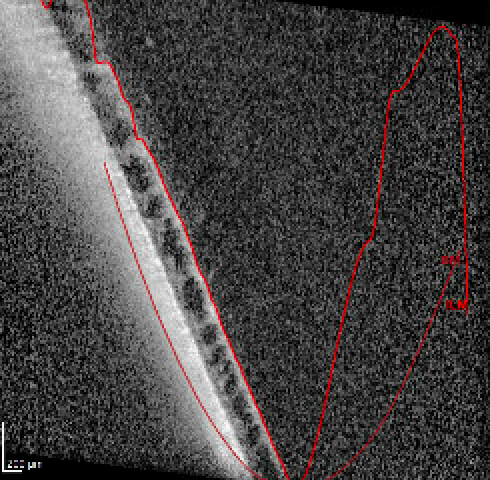
I noted that there was some peripheral schisis temporally OD that may correspond with his scotoma, though per discussion with retina, this looks like normal anatomic peripheral schisis. The scotoma seems to correspond with the area in the temporal macula/midperiphery where the retinal vein has delayed filling, but there are no other signs of BRVO. No other retinal or nerve signs of ischemia either (no PAMM, no disc edema, no RAO etc).
Here's the best quality OCT I could get through that area of temporal retina. Mac OCT was normal.
I sent a broader hypercoaguablity and inflammatory work up in addition to some labs that neurology sent from the hospital. Thus far:
Negative/normal: ESR <1, CRP <1, CBC, plts 211, RF, ANCA screening, Lyme, SPEP, homocysteine, fibrinogen, lupus anticoagulant, protein S&C, ATIII activity, APTT, PT/INR, folate, B12, trep Ab, ANA screen,
Abnormal: A1c 5.9, high thrombin time (63, normal <25).
Pending: Rickettsia (other tick screen PCR was negative), quant, lysozyme, ACE, an echo
He's seeing retina again next week -- the shallow RD in the left eye was lasered and did appear to them to be a true RD rather than schisis, but they plan to re-evaluate this prior to surgery given what's going on with the right eye.
My main questions are:
-is there something I'm missing with respect to the patchy choroidal filling in terms of work up? I have low suspicion for GCA base on young age, negative inflammatory markers and ROS, as well as (thus far) lack of amaurosis. and other signs of ischemia. Would you nonetheless do a biopsy in this patient? He is not eager to do so.
-is there a unifying pathology that would cause patchy choroidal filling and focal delayed venous outflow?
Thanks so much for your thoughts.
Erin